

Dean R. Cerio, MD
- Instructor, Division of Plastic Surgery
- Department of Surgery
- University of Alabama at Birmingham School of Medicine
- Birmingham, Alabama
Although most anemias characterized by megaloblastic erythropoiesis are due to either vitamin B12 or folic acid deficiency erectile dysfunction statistics uk buy vpxl uk, there are several other causes of megaloblastic hematopoiesis erectile dysfunction at age 35 buy discount vpxl online. Occasionally it is 2 to 3 percent erectile dysfunction after age 40 purchase generic vpxl pills, but the reticulocyte production index is low erectile dysfunction at age 24 buy discount vpxl on line, a reflection of a functionally defective marrow erectile dysfunction queensland generic 1pc vpxl mastercard. It has been suggested that these abnormalities result from 261 Hematology fragmentation of the abnormal large red cells as they pass through small arterioles impotence of organic nature order vpxl 3pc with amex. As the megaloblastic anemia becomes more sever, bizarre shapes such as triangles and helmets increases proportionately. Cells size and average number of lobes in the mature granulocyte (poly) are increased. Normally no more than 1 percent of polys have six nuclear lobes, but in megaloblastic anemia many have six or more, even ten, lobes. Despite hemolysis the reticulocyte production index is reduced because of the ineffective erythropoiesis in the bone marrow. Morphologically, the megaloblastic erythropoiesis is characterized by the presence of large cells, with asynchronism between nuclear and cytoplasmic development. Vitamin B12 Since vitamin B12 is common in human diets, almost all deficiencies of vitamin B12 are a result of malabsorption. This structure is analogous to the porphyrin structure of heme, with position of the heme iron being occupied by a cobalt atom. The vitamin B12 synthesized by microbes is deposited in animal tissues, such as liver, eggs, and 263 Hematology milk, and is therefore plentiful in fish and meat products. A normal diet contains a large excess of vitamin B12 compared with daily needs (Table 17. If malabsorption of vitamin B12 occurs, it will take 2 to 5 years before body stores are exhausted and megaloblastic erythropoiesis supervenes. A 24 hour collection of urine is begun after the radioactive B12 has been ingested. Normal subjects will excrete in their urine 7 percent or more of the radioactivity taken orally, whereas patients with pernicious anemia or other causes of vitamin B12 malabsorption will excrete well less than 7 percent. Renal insufficiency or incomplete collection of urine may result in a spuriously low excretion rate. The second part of the Schilling test is performed only if the first part gives abnormal results. In part three of the Schilling test a 2-week course of antibiotic therapy with tetracycline, 250mg four times per day, is prescribed. If bacterial overgrowth was responsible for the abnormal second part of the Schilling test, then tetracycline treatment should normalize vitamin B12 absorption. Vitamin B12 deficiency the deficiency is usually due to pernicious anemia (Table 17. Much less commonly the deficiency may be caused by veganism in which the diet lacks B12 (usually in Hindu Indians), gastrectomy or small intestinal lesions. There is no syndrome of B12 deficiency due to increased utilization or loss of the vitamin, so the deficiency inevitably takes at least 2 years to develop, i. Folic acid the terms folic acid and folate refer to a large group of compounds consisting of three moieties, pteridine, paraaminobenzoic acid, and a variable number of glutamic acid units. Folates are widely distributed in a variety of food, including green vegetables, liver, kidney, and dairy products (Table 17. During the process of intestinal absorption the folates are converted to 5-methyltetrahydrofolate, which is the main transport and storage for of folate in man. For this reason it takes 3 to 6 months for 269 Hematology tissue stores to be completely exhausted in the absence of folate replacement. Folate deficiency is most often due to a poor dietary intake of folate alone or in combination with a condition of increased folate utilization or malabsorption (Table 17. Excess cell turnover of any sort, including pregnancy, is the main cause of an increased need for folate. Aplastic Anemia Aplastic (hypoplastic) anemia is defined as pancytopenia (anemia, leucopenia, and thrombocytopenia) resulting from aplasia of the bone marrow. A selective decrease in red cell production is referred to as pure red cell aplasia. Patients with aplastic anemia generally have symptoms characteristic of a particular cellular deficiency. Those with anemia may be fatigued or short of breath, those with neutropenia may manifest serious infection, and those with thrombocytopenia may demonstrate petechiae or bleeding. A low reticulocyte count suggests underproduction rather than increased loss or destruction of red cells. The diagnosis is confirmed with a bone marrow biopsy that shows a substantial decrease in the number of red cell, white cell, and platelet precursors, and replacement of the usually cellular bone marrow with fat. Aplastic anemia can be mild or severe, and the 272 Hematology management of the patient depends on the severity of the illness. Failure of the pluripotential stem cells of the bone marrow to maintain bone marrow cellularity and the production of normal numbers of mature red cells, neutrophils, and platelets characterizes aplastic anemia. Failure of the pluripotential stem cell can be caused by many different factors (Table 17. Many agents that cause aplastic anemia, such as benzene and radiation, can on occasion precipitate malignant transformation of the damaged bone marrow stem cells, resulting in the development of acute leukemia. Pure Red cell Aplasia Acquired pure red cell aplasia is a rare disorder, usually immunologically mediated, in which there is a specific failure of production of red cells. The bone marrow biopsy shows a selective absence of red blood cell precursors, whereas white cell and platelet precursors are present in normal numbers. Anemia of Renal Failure Patients with significant renal disease almost always have anemia. Patients who require dialysis are almost always severely anemic and need repeated transfusions. The primary cause of the anemia is a lack of erythropoietin, a hormone necessary for red cell growth and development in the bone marrow. The anemia is usually normocytic and normochromic with a normal reticulocyte percentage. About 40 percent of the time, the anemia is microcytic and hypochromic, usually only mildly so, but occasionally sufficient to cause confusion with iron deficiency anemia. Inspection of the bone marrow usually shows abundant iron in reticuloendothelial cells, but little or no iron in red cell precursors. Thus, the patient has adequate iron stores, but is unable to transfer iron from the reticuloendothelial system storage cells to the red cell precursors that need it to form hemoglobin. The cause of this block in iron reutilization is uncertain, and there is no effective treatment other than to correct the 276 Hematology underlying chronic disease. Myelophthisic anemia Neoplasms, granulomatous infections, or a fibrotic process can directly replace the bone marrow. Anemias Associated with Endocrine Abnormalities [Hypothyroidism, Hypopituitarism] A mild anemia is commonly associated with hypothyroidism. The reticulocyte count is low, demonstrating that this is a hypoproliferative anemia, through the actual mechanism is not known. In hemolytic disorders, red cells are destroyed prematurely, usually in a random fashion. If the red blood cell life span is only moderately shortened, the patient will usually have little, if any, anemia because the bone marrow is capable of increasing the rate of new red blood cell production by a factor of 4 to 8. Red cell metabolism gradually deteriorates as enzymes are degraded and not replaced, until the cells become non-viable, but the exact reason why the red cells die is obscure. The breakdown of red cells liberates iron for recirculation via plasma transferrin to marrow erythroblasts, and protoporphyrin which is broken down to bilirubin. This circulates to the liver 278 Hematology where it is conjugated to glucuronides which are excreted into the gut via bile and converted to stercobilinogen and stercobilin (excreted in feces). Stercobilinogen and stercobilin are partly reabsorbed and excreted in urine as urobilinogen and urobilin. Globin chains are broken down to amino acids which are reutilized for general protein synthesis in the body. Intravascular hemolysis (breakdown of red cells within blood vessels) plays little or no part in normal red cell destruction. Extravascular Versus intravascular hemolysis There are two general sites in which hemolysis may take place (Table 17. In intravascular hemolysis, which is uncommon, red blood cells are destroyed directly within the circulatory system. Extravascular hemolysis is more common than intravascular hemolysis and involves the destruction of red blood cells within mononuclearphagocytic cells, often in the spleen. They are usually inherited, and generally (but not always) the abnormality is observable in the peripheral blood smear. Extracorpuscular defects refer to problems in the environment of the red blood cell, not in the red blood cell itself (Table 17. Extracorpuscular hemolysis is usually acquired and is often but not always discernible in the form of morphologic abnormalities in the peripheral blood smear. For example, many spherocytes suggest hereditary spherocytosis or immunohemolytic anemia and sickle cells suggest one of the sick cell syndromes. In the majority of cases hematocrit levels are normal or near normal with minimal hemolysis; greater than 25 percent (often 75%) of red cells are elliptocytes. It should be noted that some elliptical cells also occur in thalassemia, iron deficiency, myelophthisic anemias, sickle cell disease, and megaloblastic anemia. These disorders, however, are accompanied by other characteristic morphologic changes as well. Erythrocyte enzyme deficiencies Hereditary hemolytic anemia has been associated with 284 Hematology at least ten red cell enzyme deficiencies. Because of the X-linkage, male patients are more severely affected than female patients. The deficiency is not limited to any particular racial or geographically defined population. Rather than producing acute hemolysis in association with drug ingestion, it causes a chronic congenital nonspherocytic hemolytic anemia. Generally the term hemoglobinopathy is used to signify a structurally abnormal hemoglobin with at least one amino acid substitution. Structural abnormalities may cause premature red cell destruction; easily denatured hemoglobins; hemoglobins with abnormal oxygen affinity; altered hemoglobin solubility; and, in a few instances, reduced globin synthesis. In this topic only the few clinically significant hemoglobinopathies are discussed. Hemoglobin S By far the most important hemoglobinopathies are those related to the presence of sickle hemoglobin (HbS). Sickle hemoglobin results form replacement 287 Hematology of the sixth amino acid form the N-terminal end of the fichain, glutamic acid, by valine. Invariably sickle cells are typically seen on Wright-stained peripheral blood smears from patients. Hemoglobin C syndromes Hemoglobin C (HbC) is probably the second most common hemoglobinopathy (2-3% gene frequency in black populations). HbC is caused by substitution of lysine for glutamic acid in the sixth position form the Nterminal end of the fi-hemoglobin chain (same location as the substitution in HbS). A variety of acquired clinical conditions result in shortened survival of previously normal red cells. These include immune-mediated destruction, red cell fragmentation disorders, acquired membrane defects, splenic effects, and the results of infections and environmental toxins. Immunohemolytic anemia 288 Hematology Immunohemolytic anemias are the result of the binding of antibody, complement, or antibody plus complement to red cells. Antibodies formed against erythrocyte antigens may be either warm (active at 37oC) or cold (active at room temperature and below). In some cases, these antibodies activate a series of proteins, referred to collectively as complement; in others, the red cells are coated with antibody alone. As a result of complement activation by hemolytic antibodies, intravascular red cell lysis and release of hemoglobin may occur. The red cells are usually coated with IgG alone, IgG and complement or complement alone, but a minority of cases show IgA or IgM coating alone or combined with IgG antibody. Part of the coated membrane is lost so the cell becomes 290 Hematology progressively more spherical to maintain the same volume and is ultimately prematurely destroyed, usually predominantly in the spleen. The disease may occur at any age in either sex and presents as a hemolytic anemia of varying severity. Laboratory findings the hematological and biochemical finding are typical of a hemolytic anemia with spherocytosis prominent in the peripheral blood. The antibodies both on the cell surface and free in serum are best detected at 37oC. In these syndromes the autoantibody, whether monoclonal (as in the idiopathic cold hemeagglutinin syndrome or associated with 291 Hematology lymphoprolifertative disorders) or polyclonal (as following infection.
We now use recombinant human thromboplastin which is supplemented with a high concentration of phospholipid erectile dysfunction medications over the counter purchase vpxl overnight delivery. Recombinant thromboplastins are often called high sensitivity thromboplastins because they are very sensitive to reductions in vitamin K-dependent coagulation factors vasculogenic erectile dysfunction causes purchase vpxl 12pc on-line. As a consequence erectile dysfunction treatment in dubai generic 1pc vpxl visa, these thromboplastins are similar in sensitivity to the World Health Organization reference standard thromboplastin and thus have a low International Standardized Index male erectile dysfunction statistics buy cheap vpxl 9pc on-line, generally from 0 impotence natural treatment buy generic vpxl pills. Our current recombinant thromboplastin is also insensitive to therapeutic concentrations of heparin (0 erectile dysfunction treatment canada cheap 6pc vpxl with amex. In addition, this thromboplastin is generally not affected by most antiphospholipid antibodies. While many hospitals, including ours, use point of care devices to monitor warfarin, I personally am not enthusiastic about this approach. Too many preanalytical variables can affect the results, and personnel not trained in the laboratory sciences perform the test in the clinic are not necessarily the most capable in trouble shooting problems or in addressing quality assurance issues. At least recently the point of care warfarin meters have started to use high sensitivity thromboplastin, including Innovin. Other conditions (poor nutritional state, congestive heart failure, hepatic disease, hyperthyroidism, fever, steatorrhea, renal failure, antibiotic therapy, prolonged use of narcotics, etc. Other drugs and conditions (hypothyroidism, hyperlipidemia, edema, diarrhea, total parenteral nutrition, hereditary resistance to warfarin, etc. We have used Innovin from Siemens for our prothrombin testing for the last 10 years. This particular thromboplastin is insensitive to heparin, and to most antiphospholipid antibodies. This mixture triggers activation of the intrinsic pathway and subsequently, the common pathway of coagulation. The difference in test results (in seconds) before and after plasma treatment with Hepzyme is used to identify the presence of heparin in a sample. Mixing studies are based on two principles: 1) the inhibitor is in excess, and if present, it will inhibit normal and patient plasma, and 2) that 50% of any factor is enough to yield a normal test result. In a 1:1 mix, an equal volume of patient and normal plasma are mixed; in a 4:1 mix, 3 parts patient plasma is mixed with 1 part normal plasma. Depending upon the results, additional studies are performed to identify a specific factor deficiency. In normal blood coagulation, fibrinogen is converted to fibrin by the enzyme, thrombin. The first step is the thrombin-mediated proteolysis of fibrinopeptides A and B from fibrinogen. The formation of the fibrin polymer is recognized in the laboratory as the clotting time end point of the reaction. The fibrinogen value is determined from a standard curve generated by testing known concentrations of fibrinogen. In patient suspected of having a dysfibrinogen, an immunologic assay is essential to document the discrepancy between fibrinogen protein levels and function. Dysfibrinogens are characterized by the production of normal or slightly reduced levels of fibrinogen that are functionally abnormal such that tests of fibrinogen functions. Factors affecting test results (false positives and negatives) (Fibrinogen): High concentrations of heparin or fibrin degradation products can result in abnormal results. Diagnosis of hereditary fibrinogen deficiencies (both aand hypofibrinogenemia and dysfibrinogenemia), in conjunction with fibrinogen antigen and activity assays. Detection of Heparin effect 4 Detection of direct thrombin inhibitor effect, such as dabigatransee anticoagulation section on dabigatran levels. The Thrombin clotting time may be used as a qualitative measure of the level of functional fibrinogen. Prolonged times may be indicative of either decreased or markedly increased levels of functional fibrinogen; dysfibrinogenemia, or increased levels of certain fibrinogen/fibrin degradation products, heparin or dabigatran. Possible Results and Interpretation (Thrombin Time) A prolonged Thrombin Time generally means there is a reduction in the amount or function of fibrinogen. This result can occur because of inherited quantitative defects in fibrinogen production (a-fibrinogenemia or hypofibrinogenemia) or inherited qualitative defects in fibrinogen that result in the production of a normal amount (dysfibrinogenemia) or reduced amount of a dysfunctional fibrinogen (hypo-dysfibrinogenemia). Acquired dysfibrinogenemia is common in patients with severe chronic liver disease in which abnormally glycosylated forms of fibrinogen are produced. Thrombin inhibitors such as those seen after exposure to bovine thrombin or antithrombin-like substances occasionally identified in myeloma proteins or fibrin split products can also result in a prolonged thrombin time. Antithrombotic agents such as heparin, dabigatran, recombinant tissue plasminogen activator can result in an abnormally prolonged thrombin time. Factors affecting test results (false positives and negatives) (Thrombin Time): the Thrombin Time is markedly prolonged in the presence of heparin or direct thrombin inhibitors such as dabigatran. Identify heparin or a heparin-like inhibitor as the cause of a prolonged thrombin time 2. Test Principle (Reptilase Time): Plasma is incubated with Reptilase and the time to clot formation is measured. Reptilase is a thrombin-like enzyme that is derived from the venom of the South American pit viper, Fer-de-lance or the common lancehead (species Bothrops atrox). This enzyme differs from thrombin in its specificity and the extent of cleavage of the fibrinogen molecule. Reptilase cleaves only fibrinopeptide A, whereas thrombin cleaves fibrinopeptides A and B. Possible results and interpretation(Reptilase Time): A prolonged Reptilase Time may be due to decreased fibrinogen levels, dysfibrinogenemia, fibrin degradation products, soluble fibrin monomer complexes, abnormal anti-thrombins. Factors affecting test results (false positives and negatives) (Reptilase Time): Partially clotted samples (micro-clots) will cause misleading results. Under the action of thrombin, fibrinogen is cleaved to give rise to fibrin monomers. Plasmin is formed at the site of the fibrin clot attacking the fibrin clot and fibrinogen. The rapid quantitative D-dimer assay (Innovance D Dimer, Siemens, Marburg Germany) uses an enhanced latex turbidimetric method to quantify D dimer in plasma samples. Polystyrene particles with a monoclonal antibody against the D-dimer fragment covalently linked to their surface are added to the plasma specimen to be analyzed. Since the epitope against which the antibodies are directed is present twice in the D dimer fragments, only one antibody is required to cause the latex particles to agglutinate. Aggregation of the latex particles increases the turbidity of the sample allowing quantification of the D dimer fragment using the Siemens Coagulation Analyzer. D dimers are fragments of cross-linked fibrin that are produced when fibrin clot is digested by plasmin. It is important to remember that D dimer levels are a reflection of not only clot dissolution but also plasma clearance by the liver. Most d-dimer assays have been standardized to have the same cutoff value for exclusion of thrombosis. The following are two well known probability assessment scoring systems: Possible results and Interpretation (Diagnosis of Venous Thromboembolism): the D-Dimer level is calculated by the analyzer in mg/L using a standard curve. Factors affecting test results (false positives and negatives) Diagnosis of Venous Thromboembolism): Levels of plasma lipids greater than 120mg/dL, bilirubin greater than 24 mg/dl, heparin greater than 2. Plasma samples containing heterophil antibodies or mouse monoclonal antibodies used for diagnosis or therapy may lead to inaccurate results. It remains unclear whether human anti-mouse antibodies will interfere with the assay. Fibrinogen degradation fragment D will not interfere with assay in levels up to 20mg/L. Test Principle (Cryofibrinogen): Cryofibrinogens are complexes of fibrinogen, fibrin split products, and plasma globulins, that are precipitable in the cold and can be associated with purpura thrombosis, and/or hemorrhage. The plasma and blood must be separated at 37oC using heated lab equipment and rapid processing techniques. One half of the plasma is maintained in a 37oC heating block for 24 hours and the other half is placed on ice in the refrigerator for 24 hours. At the end of 24 hours, if the iced sample has gel fibrin-like strands and the 37oC does not, the sample is considered positive for cryofibrinogen. The results may be further confirmed by placing the iced sample in a 37oC incubator for 30 minutes and the gel strands should disappear, confirming the presence of a cryofibrinogen. The ansence of this cold precipitable substance in serum provides substantiation that the protein is cyrofibrinogen rather than cyroglobulin. Possible results and interpretation (Cryofibrinogen): Patient samples are scored as positive or negative. Factors affecting test results (false positives and negatives) (Cryofibrinogen): Sample must be maintained at room temperature until plasma and red blood cells are separated or the cryofibrinogen may deposit on red blood cells and give a false negative result. The activated partial thromboplastin time is the coagulation test that is used to measure the functional levels of the coagulation factors in the intrinsic pathway. Analysis is performed by making serial dilutions of the reference plasma in buffer (with assay values for the factor that needs analysis). The results are plotted on a log/log graph (polynomial curve) with percent factor on the abscissa and the time in seconds on the ordinate. The test results of the above dilutions will form a straight line that forms the basis for a standard curve at the connected points which can then be used to determine the activity of that coagulation factor in patient samples. The percent activity is read from the abscissa of the graph by finding where the clotting time obtained for the unknown sample intercepts the standard curve. All of this now is in fact programed into our analyzers, and graph paper can no longer be found in our laboratory. Isolated deficiency of Factor V activity may be inherited (incidence 1/1,000,000) or rarely acquired in patients suffering from amyloidosis. Cases of congenital factor V deficiency associated with the production of a dysfunctional molecule have also been reported. Inhibitors directed against factor V can be seen in patients exposed to fibrin glue prepared with bovine thrombin. Note: Factor V deficiency is not synonymous with Factor V Leiden (see hypercoagulable state discussion). Acquired factor X deficiency is most commonly seen in conjunction with vitamin K deficiency or therapy with vitamin K antagonists. Factors affecting test results (false positives and negatives) (Factor Assays Studies): Factor activity results can be influenced by the type of coagulometer or the reagents used to perform the test. Dilution errors can adversely affect the obtained factor activity results as well. Diagnosis of deficiency states or inhibitors affecting factors in the intrinsic pathway of the coagulation cascade. Analysis is performed by making serial dilutions (1:5 to 1:320 dilutions of calibration reference plasma or factor assay reference plasma in buffer (both with known assay values for the factor of interest). The results are plotted on a log/log graph (polynomial curve) with the percent dilution on the abscissa and the time in seconds on the ordinate. The connected points from the above dilutions form a straight line that is the basis for a standard curve. This standard curve is then used to measure the factor activity of patient samples with unknown factor content. To analyze the factor activity level on an unknown sample, 1:5 (100% activity) and 1:10 (50% activity) dilutions are prepared and mixed with immunoabsorbed, factor deficient (<1%) plasma. The percent factor activity of the patient sample is read from the abscissa of the standard curve graph by finding where the time obtained on the unknown intercepts the standard curve. Hemophilia A is inherited in an X-linked recessive manner and occurs in approximately 1 in every 10,000 live male births. These autoantibodies are associated with autoimmune disorders, malignancies, medications and the peripartum period. Like hemophilia A, hemophilia B is inherited in an X-linked recessive fashion but its incidence is about 10-fold less common occurring in 1 out of every 50-100,000 male births. These antibodies are associated with anaphylactic reactions among hemophilia B patients when receiving factor replacement. The urea stability test is sensitive to levels of 1-5%, while the acetic acid method is sensitive to below 10%. There are other assays available but only the Berichrom is approved in the United States. Of note there are reports of the thromboelastogram being abnormal, with reduced maximum amplitude and strength and increased clot cyis at 30 minutes. By comparing the difference in factor activity of the patient incubation mixture and a control mixture, the amount of inhibitor present is calculated in Bethesda units. A modification of the Bethesda assay, the Nijmegen modification, dilutes patient plasma in buffered plasma to eliminate the decrease in factor activity that occurs with pH shifts during incubation. The imprecision introduced with unbuffered plasma is relevant particularly for lower titer inhibitors. Inhibitors to specific coagulation factors can be classified as being either autoantibodies or alloantibodies. Immunosuppressive therapy is effective in eradication of these antibodies in 60-70% of patients. In contrast to autoantibodies, alloantibodies develop in patients who lack a particular coagulation factor and generate an alloantibody upon exposure to this factor during treatment with coagulation factor concentrates.
Palliation (stable disease to shrinkage) increased as needed erectile dysfunction treatment in kuwait 1pc vpxl with mastercard, if tachycardia persists erectile dysfunction symptoms order generic vpxl online. Blood pressure can be sixth month of gestation doctor for erectile dysfunction in mumbai purchase generic vpxl on-line, is possible and can be followed labile during surgery erectile dysfunction hotline order 6pc vpxl overnight delivery, particularly at the onset of intubaby uneventful childbirth erectile dysfunction funny images discount vpxl 1pc mastercard. Nitroprusside with inherited pheochromocytomas provides an opporinfusion is useful for intraoperative hypertensive crises erectile dysfunction tulsa order line vpxl, tunity to identify and remove asymptomatic tumors in and hypotension usually responds to volume infusion. It may be possible to preserve the normal the mean age at diagnosis is ~15 years lower in patients adrenal cortex, particularly in hereditary disorders in with inherited syndromes compared to those with spowhich bilateral pheochromocytomas are more likely. The diagnosis of malignant the most well-known pheochromocytoma-associated pheochromocytoma is problematic. Source: Adapted from the Freiburg International Pheochromocytoma and Paraganglioma Registry. Given the relatively high prevalence of ponent in the Krebs cycle and the mitochondrial elecfamilial syndromes among patients who present with tron transport chain. About a third of the cerebellum, kidney, brainstem, or spinal cord, sugthe patients develop metastases. About 85% of the tumors are benign nonfunctional adenomas and are usuMalignant tumors of the adrenal gland are very rare, ~1 ally discovered incidentally after abdominal imaging for case per 1 million people per year. Such tumors are called nomas may occur at any age, but two peaks are noted: adrenal incidentalomas. Arrows point to the strating tumor uptake in the right jugular glomus, the right paraganglial tumors. The bars depict the frequency of sporadic or various inherited forms 30% of pheochromocytoma in different age 20% groups. The inherited disorders are much more common among younger 10% individuals presenting with pheochro0% mocytoma. Adrenocortical carcinomas occur at Adrenocortical cancers may present in three major ways: a tenfold higher frequency among the children of southern (1) with symptoms of either glucocorticoid (or less comBrazil. Sixty percent of adrenocortical carcinomulation of genetic lesions in a fashion analogous to mas are functional (hormone secreting). Ten percent each have hyperaldosteroat that site most frequently involved in cancer. Activating mutations in catenin are Adrenocortical carcinomas may be locally invasive also noted. The European Network for the Study of threshold for performing surgery is probably too high, Adrenal Tumors proposed the following scheme based and some cancers are likely escaping early detection. Blood tests should at least one of the following factors: tumor infiltration of include fasting blood glucose, serum potassium, serum surrounding tissues (T3), tumor invasion into tumor estradiol, estrone, and adrenal androgens. In addition, a thrombus in the inferior vena cava or renal vein (T4), 24-h urine free cortisol measurement should be obtained. The prognosis is with 1 mg of dexamethasone given at bedtime and serum related to disease stage. Imaging is a critical tool in assessing the benign or malignant nature of an adrenal mass. Benign tumors are also as adjuvant therapy for at least 2 years or until disease homogeneous with a smooth border. Although useful distinguishing feature is the speed with which response may be noted from mitotane or mitotane comcontrast material is washed out of the mass. Benign bined with other chemotherapeutic agents, responses are masses tend to show rapid contrast washout; more than often short and the survival of patients has not been half the contrast is gone 10 min after administration. Chemical shift imaging some palliation, although it would be best if patients can distinguish the protons in fat from those in water; the with this unusual cancer were part of organized clinical lipid content of tumors is usually lower than that of adetrials. If the imaging on mitotane should be treated to prevent hypocortitests provide cause for concern based on size (>4 cm), solism (preferably with prednisone) and monitored for the contour (irregular), inhomogeneity, density, and slow development of hypoaldosteronism. In the setting of contrast washout, the definitive approach to obtaining incomplete resection or recurrence of functional tumors, tissue is a surgical procedure in which the adrenal gland hypercortisolism may be managed ketoconazole is excised in its entirety. Doses of the antagonists may need to be increased Pract 14:1137, 2008 based on the adrenal suppression noted in each individual. J Clin Endocrinol Metab 92:1217, 2007 Terzolo M et al: Management of adrenal incidentaloma. Paraneoplastic syndromes refer to Etiology the disorders that accompany benign or malignant tumors but are not directly related to mass effects or Hormones can be produced from eutopic or ectopic invasion. Many hormones are produced at low levels from a paraneoplastic syndromes indicate that they are more wide array of tissues, in addition to the classic endocrine common than is generally appreciated. Thus ectopic expression is often a quantitative toms, and metabolic alterations associated with paranechange rather than an absolute change in tissue expresoplastic disorders may be overlooked in the context of a sion. Consequently, atypical entrenched and conveys the abnormal physiology associclinical manifestations in a patient with cancer should ated with neoplastic hormone production. The high levels of hormones, ectopic expression is typically most common endocrinologic and hematologic syncharacterized by abnormal regulation of hormone prodromes associated with underlying neoplasia are disduction. It is likely that cellular dedifferentiation underlies esophagus, breast, genitourinary tract, and in multiple most cases of ectopic hormone production. These expression profiles are likely to be driven cellular proliferation and differentiation in other tissues by alterations in transcriptional repression, changes in including skin, bone marrow, breast, and hair follicles. Thus abnormal expression of these develduction can be stimulated by mutations in oncogenes, opmental transcription factors appears to provide a link by altered expression of viral or cellular transcription between cell proliferation and differentiation. Moreover, the dihydroxyvitamin D, leading to enhanced gastrointestinal paraneoplastic endocrinopathies are sometimes the precalcium absorption. For most ectopic hormone syndromes, an extensive list of tumors has been reported to produce one or more hormones. The mechanism of activation of the vasopressin gene levels may be increased in patients with lymphoma. Saline rehydration is used to symptoms refiects the rapidity of onset as well as the dilute serum calcium and promote kaliuresis. Bisphosphonate infuthe setting of an inappropriately normal or increased sions can be repeated or oral bisphosphonates can be urine osmolality. Other ered in severe hypercalcemia when saline hydration and causes of hyponatremia should be excluded, including bisphosphonate treatments are not possible or are too renal, adrenal, or thyroid insufficiency. Hypercalcemia associshould also be considered as possible causes of hyponaated with lymphomas, multiple myeloma, or leukemia tremia. Vasopressin assay is not usually necessary to make may respond to glucocorticoid treatment. The disorder should be corrected gradually unless menVasopressin is an antidiuretic hormone normally produced tal status is altered or there is risk of seizures. Compensatory insensible losses, is often sufficient to partially correct mechanisms, such as decreased thirst, suppression of aldoshyponatremia. The rate of sodium correction centripetal fat redistribution, probably because the should be slow (0. However, because this product activity through the mineralocorticoid receptor, leading lacks the signal sequence necessary for protein processto severe hypokalemia. Patients may experience In most cases, the tumor causing hypoglycemia is depression or personality changes because of extreme clinically apparent and hypoglycemia develops in associcortisol excess. The diagnosis is made by documentbetes mellitus and hypokalemia can worsen fatigue. Poor ing low serum glucose and suppressed insulin levels in wound healing and predisposition to infections can association with symptoms of hypoglycemia. Treatment of the underlying malignancy, if underlying malignancy, measures to reduce cortisol levpossible, may reduce the predisposition to hypoels are often indicated. Glucocorticoid replacement should be extragonadal germinomas, lung cancer, hepatoma, and provided to avoid adrenal insufficiency. Treatment should be directed at the underlying some 11p15 locus that is normally imprinted (that is, malignancy. Treatment involves Gastrointestinal removal of the tumor, if possible, and supplementation cancer with phosphate and vitamin D. Octreotide treatment Breast cancer reduces phosphate wasting in some patients with tumors Genitourinary cancer that express somatostatin receptor subtype 2. The elevation of granulocyte, platelet, and eosinophil counts in most patients with myeloproliferative disorders detected on a routine blood count. Approximately 3% of is caused by the proliferation of the myeloid elements patients with renal cell cancer, 10% of patients with due to the underlying disease rather than a paraneoplastic hepatoma, and 15% of patients with cerebellar hemansyndrome. In most cases the eryin patients with solid tumors are less well characterized throcytosis is asymptomatic. If the red cell mass is elevated, the serum the paraneoplastic syndromes parallels the course of the erythropoietin level should then be measured. Ectopic production of erythropoietin by cancer cells causes most paraneoplastic erythrocytosis. If the tumor cannot be resected or by cancer cells may stimulate erythropoietin release but treated effectively with radiation therapy or chemotherhave not been proven to cause erythrocytosis. The other patients have proteins in urine and serum patients with cervical, gastrointestinal, renal, and breast that stimulate the growth of bone marrow cells. However, the etiology of granulocytosis has not been characterized in most patients. Patients with granulocytosis are nearly all asymptomatic, and the differential white blood cell count does not Treatment: have a shift to immature forms of neutrophils. Patients with advanced-stage disease shortness of breath related to eosinophilia, symptoms are more likely to have granulocytosis than those with resolve with the use of oral or inhaled glucocorticoids. The etiology of Pathogenesis thrombocytosis has not been established in most cases. Patients with thrombocytosis are nearly all asymptoPatients with cancer are predisposed to thromboemmatic. Thrombocytosis is not clearly linked to thrombobolism because they are often at bedrest or immobilized, sis in patients with cancer. In addition, clot20% of patients with breast, endometrial, and ovarian ting may be promoted by release of procoagulants or cancers, and 10% of patients with lymphoma. Patients cytokines from tumor cells or associated infiammatory with thrombocytosis are more likely to have advancedcells, or by platelet adhesion or aggregation. The specific stage disease and have a poorer prognosis than patients molecules that promote thromboembolism have not without thrombocytosis. In addition to cancer causing secondary thrombosis, primary thrombophilic diseases may be associated with cancer. About 20% of patients with this syndrome Tumors and tumor cell lines from patients with lymhave cancers. The most common cantive contraindication to heparin anticoagulation (hemcers associated with thromboembolic episodes include orrhagic brain metastases or pericardial effusion) should lung, pancreatic, gastrointestinal, breast, ovarian, and genibe considered for placement of a filter in the inferior tourinary cancers, lymphomas, and brain tumors. Patients vena cava (Greenfield filter) to prevent pulmonary with cancer who undergo surgical procedures requiring embolism. Patients with cancer who undergo a major surgical procedure should be considered for heparin prophylaxis or pneumatic boots. Breast Diagnosis cancer patients undergoing chemotherapy and patients the diagnosis of deep vein thrombosis in patients with with implanted catheters should be considered for procancer is made by impedance plethysmography or bilatphylaxis (1 mg/d warfarin). If compression ultrasonography is normal and a high clinical suspicion exists for deep vein Neurologic paraneoplastic syndromes are discussed in thrombosis, venography should be done to look for a Chap. Elevation of D-dimer is not as predictive of deep vein thrombosis in patients with cancer as it is in patients without cancer. A retrospective study of 453 patients embolus need no additional tests for cancer other than a treated in a single institution in a 10-year period. Lung Cancer 2009; June 15 (Epub ahead of print) careful history and physical examination. Neuronal cell-surface antigens can be the target of antibodies in some patients with paraneoplastic encephalitis. First, it is common for Syndromes of the spinal cord Subacute necrotizing myelopathy symptoms to appear before the presence of a tumor is Motor neuron dysfunction known. Second, the neurologic syndrome can evolve Myelitis in a rapidly progressive fashion, producing a severe Stiff-person syndrome and usually irreversible neurologic deficit in a short Syndromes of dorsal root ganglia period of time. Therefore, the Multiple levels of involvement a major concern of the physician is to recognize a disEncephalomyelitis, sensory neuronopathy, autonomic dysfunction order promptly as paraneoplastic in order to identify Syndromes of peripheral nerve and treat the tumor. In these cases, a biopsy Autonomic neuropathy of the affected tissue is often difficult to obtain, and Syndromes of the neuromuscular junction although useful to rule out other disorders. Common features antibody specificity often correlating with the tumor of these four antibodies are that they target cell-surface type. These include several syncancer, particularly metastatic and leptomeningeal disdromes of infiammatory neuropathies and myopathies.
Cy5 dye a proprietary name for a sulfoindocyanine dye having kmax these are transported to tissues sublingual erectile dysfunction pills purchase cheap vpxl on line, which convert them to the cytoexcitation 649 nm and kmax emission 670 nm erectile dysfunction for young men buy discount vpxl online, and uses similar to toxic agent yohimbine treatment erectile dysfunction purchase vpxl on line amex, phosphoramine mustard erectile dysfunction drugs at walmart purchase vpxl toronto, and acrolein erectile dysfunction bp meds cheap vpxl 3pc overnight delivery. It is usually benign erectile dysfunction gene therapy treatment order vpxl master card, pyridoxine-responsive, and the secretion of gonadotropins and spermatogenesis in humans. Cyr61 a growth factor binding protein expressed from G0/G1 to midcystatin any of a group of proteins, present in tissues and body fluG1 of the cell cycle. Family 3 proteins (also called kininogens) contain three cysit represents L-half-cystine. Examples: cystatin A (also prising a central cavity lined with epithelium and containing fluid called stefin A); type 2 cystatin C (also called neuroendocrine basic or semisolid material. They are commonly encountered during sequencing of peptides and proteins as the end product O H N H of oxidative cleavage of the disulfide bonds of cystine residues and 2 S concomitant oxidation of cysteine residues. The enzyme carries out the final reaction in cysteine the trivial name for b-mercaptoalanine; a-amino-b-thiol2 the pathway for the synthesis of cysteine from methionine. In neutral or alkaline sohydro-lyase (adding homocysteine); other names: serine sulfhylution it is readily oxidized by air to cystine. This is the first step in the synthesis of mammals it is a nonessential dietary amino acid and is glucogenic. An enzyme in the pathway for the their stereochemical designation as either R or S being the converse synthesis of methionine in plants and bacteria. It catalyses the forof that possessed by most chiral a-amino acids found in proteins. It was characterized by having a cysteine residue at the active centre and originally found in transforming growth factor-fi and platelet-deby being irreversibly inhibited by sulfhydryl reagents such as iodoacetate. These are cystinosin a membrane protein of lysosomes that is encoded by the (1) papain-like proteases; (2) caspase-hemoglobinase proteases; and gene for cystinosis. See cathepsin B, cathepsin H, cathepsin segments, and 8 potential glycosylation sites. Without treatment the result is end-stage renal failure minal amino-acid residue from a protein. There heterozygotes have abnormal urinary excretion of cystine and basic is an abnormality in the secretions of the exocrine glands, which amino acids. It is converted metabolically to 1-b-D-arabinofuranosyling of a chloride ion channel. As it is somewhat insoluble in neutral aqueous solutions, it sometimes forms urinary stones. This diester then readily hydrolyses to a mixture of the two monoesters cytidine 2fi,3fi-(cyclic)phosphate see cytidine phosphate. N It participates as coenzyme in reactions in which a cytidylyl group is transferred. The position of the phosphoric residue on the ribose moiety of a given ester may be specified by a prefixed locant. It is reduced by cycytidylyl the cytidine[mono]phospho group, the acyl group derived tochrome b5 reductase. The It is cytosolic in erythrocytes and a myristoylated peripheral memmost notable are inhibition of cytoplasmic cleavage following nubrane protein of 275 amino acids in hepatocytes. Point mutations in clear division, induction of nuclear extrusion, and inhibition of cell the gene cause hereditary enzymopenic methemoglobinemia. They bind to the growing plus ends of actin filaments, precytochrome b6 or cytochrome b563 a b-type cytochrome that conventing further addition of actin molecules, and thus affect functains two heme moieties and forms part of cytochrome b6f complex tions that involve assembly and disassembly of actin filaments. The protein is a target cell, appropriate to the physiological effect of the hormone in dimer (human) of a (the glycoprotein gp91-phox, which contains question, by quantitative cytochemical methods. X-linked chronic granulomatous disease, while similar defects in cytochemistry the chemistry of living cells, especially the cellular the b subunit cause an autosomal recessive form of the same dislocalization of biochemical substances and processes; microscopical ease. The sequence from many (>100) species has been maining members are designated by a suffixed number based upon determined. About 26 residues are completely invariant, especially the wavelength (in nanometres) of the a-band. It is released into the cytosol after a formyl group) as prosthetic group, not covalently bound to the induction of apoptosis, binds to Apaf1, and activates procaspase 9. It transfers electrons directly to cyCytochromes d contain a tetrapyrrollic chelate of iron as prosthetic tochrome c. The reaction is: a-band, b-band, and c-band (or Soret band), resulting from the ab4 ferrocytochrome c + O2 = 4 ferricytochrome c + 2 H2O. Elec(b), 531 (c), and 524 (c1) nm; and for the c-band 439, 429, 415, and trons originating in cytochrome c are transferred via heme a and 418 nm. Subunit I cytochrome b a component of the eukaryotic mitochondrial respibinds the two hemes and CuB. Several mutations in cytochrome f or cytochrome b552 a cytochrome, designated after the it result in mitochondrial myopathy or cardiomyopathy. Cytochrome b2, is a mitochondrial intermembrane-space cytochrome h a b-type cytochrome found in the hepatopancreas of protein induced by lactate during respiratory adaptation (in yeast). Cytokines elicit a variety of responses cytochrome o any b-type prokaryotic cytochrome containing prodepending on the cytokine and target cell. Their actions include toheme as the prosthetic group that, unlike a typical cytochrome b, control of cell proliferation and differentiation, regulation of imserves as a terminal electron acceptor (cytochrome oxidase) and is mune responses, hemopoiesis, and inflammatory responses. See also angiogenin, epidermal growth factor, erythropoietin, fibrobabsorption peak (Soret band) near 450 nm. In mammalian liver a stimulatory effect on the division of plant cells and a retarding efmany cytochromes P450 are inducible by drug and other treatment, fect on leaf senescence. They are classed cytology the branch of science dealing with the origin, structure, into 27 families, of which 10 are found in all mammals. It includes the consists of a group of proteins that exhibit 40% or greater sequence (microscopic) examination of cells, particularly in the detection and identity, and the family is described by an Arabic number. Its N-terminal region is hydrophobic enclosed in a cell-derived lipid membrane envelope containing virus and keeps the enzyme attached to the endoplasmic reticulum, while encoded glycoproteins that are important for the attachment of the the rest of the protein projects into the cytosol. In some cases these cyton or cytone the body of a cell, especially a nerve cell, as distinct pigments were subsequently shown to be riboflavin or a flavin-confrom its processes. The technique has many applications, including specific droplets, molecules, or particles are transported across a polarized labelling of cells using fluorescent antibodies, using fluorescent cell from one surface to another. Antibody thus herited traits with cytological features, especially the appearance, fixed to a cell is still able to bind antigens in the vicinity; i. Cytohelicase the proprietary name for a commercial enzyme prepacytophotometry a technique that employs a combination of miration consisting predominantly of a 1,3-b-D-glucanase. It is useful croscopy and spectrophotometry for the detection, localization, for the isolation of protoplasts because it lyses cell walls. It may be done on stained cytohemolysin any of a family of pore-forming toxins of molar or unstained preparations. The term is not synonymous with cytoplas+cytosis suffix indicating an increase in the number of cells (in an mic fraction. It may be further resolved into a mitochondrial functions, including cellular movement, cell division, endocytosis, fraction, a microsomal fraction, and a soluble fraction. Subsequently the cytoskeleton was rewhose determinants are not located on a chromosome. The detergarded also as a possible subcellular site of action of various horminants may be. The term is within the cytoplasm of eukaryotes, such as the rough and smooth not synonymous with soluble fraction. See also microsomal cytosol aminopeptidase another name for leucyl aminopeptidase. D600 gallopamil, 5-methoxyverapamil, a-(3[2-(3,4-dimethoxydAdo symbol for a residue of the deoxynucleoside deoxyadenosine; phenyl)ethyl] methylamino propyl)-3,4,5-trimethoxy-a-(1-methyl2fi-deoxyribosyladenine; adenine 2fi-deoxyriboside (alternative to ethyl) benzeneacetonitrile; a drug used in the laboratory to block dA). Partial loss-of-function mutations result in a long-lived used as a herbicide and to increase latex output from old rubber phenotype with resistance to killing by several bacterial pathogens. It is intro165 danazol data cleaning for 2fi-deoxyadenosine 5fi-phosphate; 2fi-deoxy-5fi-adenylic acid; adevice in the absence of incident radiation. On diverging, the light does not enter the apersteroids and acts as a ligand for their receptors. However, they are subject to regulatory O mechanisms that are stimulated by light, and inhibited by the dark. It is dephosphorylated mainly by calcineurin, and is involved in the psychomotor effects of caffeine. It acts as a competitive antagonist wavelength rhodopsin from the bumblebee (Bombus terrestris) in of p-aminobenzoic acid and an inhibitor of folate synthesis. See Duffy contain a user-specified pattern, each time refining the results via system. It is a glycoside dC symbol for a residue of the deoxynucleoside deoxycytidine (alterformed from the tetracyclic aglycon daunomycinone (C21H18O8) native to dCyd). Its N-terminal extracellular segment contains four immunoglobulin-like and seven fibronectin repeats, and shows about 30% homology with netrin receptor. Essentially it was a lamellar model: two cytidylate hydroxymethyltransferase; an enzyme that catalyses the sheets of protein separated by a sheet of lipid. To account for 5,10-methylenetetrahydrofolate + H2O + deoxycytidylate = the permeability properties of the plasma membrane the model was tetrahydrofolate + 5-hydroxymethyldeoxycytidylate. See pterin-4aacids, for which the prototype is the Rho guanine nucleotide excarbinolamine dehydratase. They are three-letter or one-letter symbols for ribonucleosides in order to form symbols for 2fi,3fi-dideoxyribonucleosides. It has a very long persistence of activity de-ashing a procedure in which interfering inorganic. Cl death domain a region of limited similarity, comprising ~80 Cl Cl residues close to the intracellular C terminus of certain cell-membrane receptors. It oligomerizes on binding of its ligand and contains an intracellular death domain that recruits a cytosolic death domain protein. They are abundant in phagocytes, in the small intestinal mucosa of decapentaplegic abbr. Degrowth factor b superfamily that triggers various morphogenetic fensins adopt multimeric pore-forming complexes in membranes, processes in Drosophila. It participates in an intercellular signalling thereby rendering the membrane permeable. They are distinguished pathway that activates a specific transcription factor required for by having a predominantly beta sheet structure. A homologue of dpp occurs in deficiency disease any disease resulting from the deficiency of one mammals. The reaction may be enof all the constituents, including trace substances, are quantitatively zyme-catalysed by a decarboxylase or, in some instances, particuknown. One such is the genetic code, wherein a specific atom or molecule from an excited state. The term is used especially in redegranulation 1 any process by which granular cells lose their grangard to microbes, toxic chemicals, carcinogens, and radioactive ules. It denotes the same interval of temperature as the kelvin on a can chain, together with other oligosaccharide substituents. A deletion can vary in size from a single nucleotide residue to dehydro+ prefix (in chemical nomenclature) denoting the loss of a segment containing a number of genes. In common usage it is often used in place of at one end of the chromosome, whereas an intercalary deletion ocdidehydro+;. The gene at 11q12-q13 encodes a multipass Fluoroimmunoassay; a collection of proprietary diagnostic methtransmembrane protein of 475 amino acids. H C O delta symbol: d (lower case) or D (upper case); the fourth letter of the 3 Greek alphabet. Compare nitrification, nitrogen fixadantly expressed in brain, heart, skeletal muscle, kidney, and lung, tion (def. It is useful for scanning chrosive loss of mineral salts from the tissues, especially bone, as occurs matograms or electrophoretograms, for determining the opacity of in certain diseases. It is sometimes taken to include disulfide density 1 a or mass density symbol: q; the mass of a substance per bond rupture or chemical modification of certain groups in the prounit volume.
India Cancer incidence in 5 Indian cities correlated to background Decreasing incidence with increasing background dose (Nambi & Soman erectile dysfunction drugs medicare cheap vpxl 1pc with mastercard, fi-radiation of 0 erectile dysfunction aafp order vpxl overnight. Several studies have been performed to derive indoor and outdoor doses erectile dysfunction diabetes causes buy vpxl 3pc overnight delivery, and individual doses have been measured with personal dosimeters erectile dysfunction age factor buy vpxl 6pc on line. More than 80 000 individuals who live in the high background areas were estimated to receive an annual dose to the red bone marrow of 2 erectile dysfunction caused by sleep apnea order vpxl line. Nonsignificantly lower rates of mortality from all cancers and from leukaemia erectile dysfunction daily medication buy vpxl on line, breast cancer and lung cancer were found in the high background areas (Tao & Wei, 1986; Wei et al. Although a significantly higher risk for cancer of the cervix uteri was found in the high background area, it was considered not to be due to the ionizing radiation. Nevertheless, a higher frequency of stable chromosomal aberrations (translocations and inversions) was found in the high-dose area (see section 4. Correlations were found only in women, with positive correlations for stomach cancer and uterine cancer and negative correlations for cancers of the breast, lung, pancreas and oesophagus. Although studies have shown very little evidence for an excess cancer risk, they have been of limited quality. Cancer incidence and background fi-radiation were investigated in five Indian cities with background levels of 0. A significantly decreased overall cancer incidence was observed with increasing dose, but the authors underlined the limited extent of cancer registration in India. Background radiation did not appear to affect the rates of leukaemia or of cancers of the breast, intestine or lung. The findings are based on few cases, and increased ascertainment and medical surveillance are likely to have influenced them. The results of surveillance of childhood leukaemia in cancer registry populations from 1980 up to the end of 1991 were reported by Parkin et al. Although there was a slight increase in the incidence of childhood leukaemia in Europe during the period studied, the overall geographical pattern of change bears no relation to estimated exposure to radiation from the Chernobyl fall-out. The incidence of leukaemia among infants in Greece after exposure in utero as a consequence of the Chernobyl accident was found to be higher in children born to mothers who lived in areas with relatively greater contamination (Petridou et al. On the basis of 12 cases diagnosed in infants under the age of one year, a statistically significant increase in the incidence of infant leukaemia was observed (rate ratio, 2. The authors concluded that the observed increase was not related to exposure to radiation from the Chernobyl accident. A cluster of childhood leukaemias was reported around the Sellafield nuclear installation in the United Kingdom in 1983 (Black, 1984). Childhood leukaemia was subsequently reported to be occurring in excess in other regions of the United Kingdom where there were nuclear installations, although the incidence of all cancers was not increased (Forman et al. More refined analyses in the United Kingdom gave little evidence that the incidence of childhood leukaemia was related to proximity to nuclear facilities, except for the Sellafield installation (Bithell et al. Another study conducted in England and Wales showed that the incidence of childhood leukaemia was increased around sites selected for nuclear facility construction but in which the facilities had not been completed (Cook-Mozaffari et al. An infectious agent associated with large migrations of people into these areas has been proposed as a possible explanation for the clusters (Kinlen et al. These ecological analyses are severely limited by the absence of information on individual doses of radiation, but they were probably lower than the dose of natural background radiation (Darby & Doll, 1987). Four cases of leukaemia were seen among children whose fathers received doses fi 10 mSv within six months of conception (Gardner et al. These findings were not replicated in a similar but smaller study at the Dounreay nuclear facility in Scotland (Urquhart et al. Further, a study of 10 363 children who were born to fathers who worked at the Sellafield facility included an evaluation of the geographical distribution in the county of Cumbria of the paternal dose received before conception. The paternal doses were consistently higher for fathers of children born outside Seascale, a village close to Sellafield where the original cluster was found. Since the incidence of childhood leukaemia was not increased in these areas of West Cumbria, despite the higher preconception exposures, the authors concluded that paternal exposure to radiation before conception is unlikely to be a causal factor in childhood leukaemia (Parker et al. A further study of cancer among the children of nuclear industry employees in the United Kingdom was conducted with a questionnaire approach (Roman et al. Employees at three nuclear establishments were contacted, and 111 cancers (28 leukaemias) were reported among 39 557 children of male employees and 8883 children of female employees. The incidences of all cancers and of leukaemia were similar to those in the general population; however, the rate of leukaemia in children whose fathers had accumulated a preconceptual dose fi 100 mSv was significantly higher (5. Further, the approach used probably resulted in substantial under-ascertainment of the number of cases of childhood cancer because no effort was made to obtain information on children of workers who had died; ex-employees who were not on the pensions database were not contacted; ex-employees of one of the three nuclear establishments and persons over the age of 75 were not contacted at all; an unstated number of questionnaires was returned undelivered; and 18% of the male workers who received the questionnaires failed to return them. Comparison with a record linkage study that included all children of nuclear industry workers in the United Kingdom (Draper et al. The study is therefore susceptible to biases related to incomplete ascertainment of children with cancer and to the reasons for responding or failing to respond to the questionnaire. Any increase in the incidence of cancer would thus be expected to be negligible and undetectable (Upton, 1981). An ecological survey found no link between estimated patterns of radiation release and increased cancer rates (Hatch et al. Other studies of the Three-Mile Island incident have given inconsistent results (Fabrikant, 1981; Wing et al. These approaches, which have drawn on information obtained from both epidemiological and experimental studies, have then been used to estimate the risks from various exposures. In this section, several measures of risk are defined, problems and uncertainties in estimating those risks are discussed, and recent efforts of major national and international groups to provide quantitative risk estimates are summarized. Several estimates of risks from particular sources of exposure are given as illustrations. An important measure of risk is the lifetime risk that an individual will die from a cancer that has been caused by exposure to a carcinogenic agent such as radiation. The lifetime risk is sometimes referred to as the risk of exposure-induced death and differs from the excess lifetime risk (National Council on Radiation Protection and Measurements, 1997), which does not include deaths from cancers that would have occurred without exposure but which occur at a younger age because of the exposure (Thomas et al. Such risks are dependent on dose and thus must be expressed as a function of dose. In the most commonly used linear model, the risk is often expressed per unit of dose. Lifetime risk estimates may depend on sex, age at exposure and the pattern of exposure over time. Approaches have been developed that allow estimation of sex-specific lifetime risks resulting from various patterns of exposure with regard to age and time. For example, the risk from single exposures at various ages or from continuous exposure over a specified period can be estimated. Lifetime risks also depend on many individual characteristics, but too little is known about such dependence for it to be taken into account. Rigorous definitions and interpretations of measures such as attributable risk and the probability of causation have been discussed extensively (Greenland & Robins, 1988). Once a model for estimating the lifetime risks of individuals has been developed, it can be applied to all individuals in a population (such as an entire country) to estimate the total number of cancers that are expected to occur as a result of exposure to various specified doses. Since risks often depend on sex and age at exposure, such estimates require demographic data on the population for which the risk estimates are being made. Like individual risks, population risks can be expressed as a function of dose, or per unit of dose for a linear model. If estimates of the doses or distribution of doses received by a population from a particular source of radiation are available, the models for estimating lifetime risks can be applied to estimating the number of cancer deaths associated with exposure from the source. This number is sometimes expressed as a fraction of the total number of cancer deaths that have occurred in the population and is known as the attributable risk. Radiation risks are commonly measured in terms of cancer mortality, but all of the above measures can also be used to estimate the risk for non-fatal cancer. An additional measure that takes account of age is the loss of life expectancy, which was defined and discussed by Thomas et al. This is sometimes expressed as the number of years of life lost per radiation-induced cancer. The most recent attempts at risk assessment are based on epidemiological data, so that age-specific cancer mortality or incidence rates are estimated as a function of baseline rates and parameters that characterize the relationship between risk and exposure to radiation. The risk from radiation is usually expressed as a function of dose, age at the time of exposure, time since exposure, sex and sometimes other factors. These functions are then used in combination with data on the characteristics of the population. A key feature of this model is that risks are expressed relative to the baseline rather than in absolute terms. If life-table methods are used, the age-specific risks fi (a, s) and fif (a, s, D, e, t) can be applied to demographic data for the population for which risk estimates are being made to obtain lifetime risk estimates or any of the other measures described above. Because the populations and exposures for which risk estimates are desired nearly always differ from those for which epidemiological data are available, assumptions are required, many of which involve considerable uncertainty. Some of the more important assumptions are discussed below, and the approaches used to address these problems in specific risk assessments are described in section 2. Most situations for which risk estimates are desired involve exposure to low doses and dose rates. Because the estimates obtained directly from epidemiological data on populations exposed to low doses are imprecise, it is necessary to extrapolate from risks estimated for persons exposed to higher doses and dose rates than those of direct interest. Specifically, the data on the atomic bomb survivors have played a strong role in developing models for risk estimation, and estimates based on those data tend to be driven by doses > 1 Gy, which is much higher than the doses for which risk estimates are needed, < 0. Although a factor of 2 has been used in several risk assessments, the magnitude of the factor, or whether it is needed at all, is uncertain. Although the risk for cancer associated with exposure to radiation has been found to depend on sex, age at exposure and the time between exposure and diagnosis or death, the available data are not adequate to determine the exact form and magnitude of such dependence, and risk estimates are usually based on relatively simple assumptions. The risks of people exposed at young ages are particularly uncertain, since follow-up of these persons is the least complete. The data on many cancer types indicate that the relative risk is greatest for people exposed early in life, but the magnitude of the increase and whether it persists throughout life is highly uncertain. Another difficulty is that the baseline cancer risks of the population being studied may differ from those of the population for which risk estimates are desired. The risk for breast cancer can be estimated from the results of studies of white women (Abrahamson et al. The problem is less severe for leukaemia and for all solid tumours combined, since the baseline rates for these categories do not vary as greatly among countries. A closely related problem is that smoking and other life-style factors may modify risks. This is especially important in estimating risks for individuals, but also affects population risks if these factors differ in the population used to develop the risk models and in that for which risk estimates are desired. In fact, these differences are probably part of the reason for differences among baseline rates in different countries. Increasing attention is being given to quantifying the uncertainties in risk estimates. The sources of uncertainty include lack of knowledge about the correct assumptions, as discussed above, and these uncertainties must often be assessed subjectively. Still other sources of uncertainty are possible errors and biases in the epidemiological data used, including errors in the estimated doses. Methods are available for addressing these uncertainties, but they are often difficult to apply and require a thorough understanding of the magnitude and nature of the errors. Estimates were given for death from leukaemia, from all cancers except leukaemia and from several other types of cancer. The lifetime risk estimates were based on demographic data for the population of Japan in 1982. Alternative estimates based on patients with ankylosing spondylitis or cervical cancer who were exposed to radiation were also given. Because baseline risks increase as persons age, the multiplicative model generally results in larger estimates than the additive model.
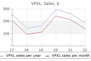
Buy vpxl 12pc with amex. Signs Of Erectile Dysfunction or Impotence - by Dr Sam Robbins.